C9: Membrane Transport
Introduction: The Cell’s Gatekeeper
Every living cell is surrounded by a cell membrane, a remarkable structure that acts as a selective barrier. It controls what enters and leaves the cell, maintaining the specific internal environment necessary for life. This regulation of passage is known as membrane transport. Understanding how this works is fundamental to understanding cell function, physiology, and even disease.
This pre-lab reading will guide you through the structure of the cell membrane and the various ways substances cross it.
The Fluid Mosaic Model: A Dynamic Barrier
Recall from previous lessons that the cell membrane is described by the fluid mosaic model. It’s primarily composed of:
- Phospholipid Bilayer: A double layer of phospholipid molecules. Their hydrophilic (water-loving) heads face the watery environments inside and outside the cell, while their hydrophobic (water-fearing) tails face inward, creating a barrier to water-soluble substances.
- Proteins: Embedded within or attached to the bilayer. These proteins play crucial roles in transport, signaling, and cell recognition. Transport proteins, in particular, are essential for moving specific substances across the membrane.
- Cholesterol: Found in animal cell membranes, cholesterol helps regulate membrane fluidity.
- Carbohydrates: Often attached to proteins (glycoproteins) or lipids (glycolipids) on the outer surface, involved in cell recognition and adhesion.
Types of Membrane Transport
Movement across the membrane can be broadly categorized based on whether the cell needs to expend energy.
1. Passive Transport: No Energy Required
Passive transport mechanisms move substances down their concentration gradient (from an area of high concentration to an area of low concentration) without the cell expending metabolic energy (like ATP).
a) Simple Diffusion
- What: Direct movement of small, nonpolar molecules (like O2, CO2, steroid hormones) across the phospholipid bilayer.
- How: Molecules dissolve in the lipid bilayer and move from high to low concentration.
- Rate Factors: Depends on the steepness of the gradient, temperature, size of the molecule, and lipid solubility.

b) Facilitated Diffusion
- What: Movement of larger polar molecules (like glucose, amino acids) or ions (like Na+, Cl-) across the membrane down their concentration gradient.
- How: Requires the help of specific transport proteins:
- Channel Proteins: Form hydrophilic pores through the membrane. Some are always open (leak channels), while others are gated (open/close in response to stimuli). Aquaporins are channel proteins specific for water.
- Carrier Proteins: Bind to the substance, change shape, and shuttle it across the membrane.
- Rate Factors: Limited by the number of available transport proteins (saturation can occur).

c) Osmosis
- What: A special case of diffusion involving the net movement of water across a selectively permeable membrane. Water moves from an area of higher water concentration (lower solute concentration) to an area of lower water concentration (higher solute concentration).
- How: Water can move slowly across the lipid bilayer or more rapidly through specific channel proteins called aquaporins.
- Tonicity: Describes the relative solute concentration of two solutions separated by a membrane.
- Isotonic: Solutions have equal solute concentrations. No net water movement.
- Hypertonic: Solution has a higher solute concentration (lower water concentration) than the other. Water moves out of the other solution/cell.
- Hypotonic: Solution has a lower solute concentration (higher water concentration) than the other. Water moves into the other solution/cell.
Effects of Tonicity on Cells:
- Animal Cells:
- Isotonic: Normal shape.
- Hypertonic: Cell shrinks (crenation) as water leaves.
- Hypotonic: Cell swells and may burst (lysis) as water enters.
- Plant Cells: Have a rigid cell wall.
- Isotonic: Cell is flaccid (limp).
- Hypertonic: Cell membrane pulls away from the cell wall (plasmolysis) as water leaves the central vacuole.
- Hypotonic: Cell becomes turgid (firm) as water enters, pushing the plasma membrane against the cell wall. This turgor pressure is essential for plant support.

Watch this video for a clear explanation of Osmosis:
2. Active Transport: Energy Required
Active transport moves substances against their concentration gradient (from low concentration to high concentration). This process requires the cell to expend energy, usually in the form of ATP (adenosine triphosphate). It always involves specific carrier proteins often called “pumps”.
a) Primary Active Transport
- How: Energy from ATP hydrolysis is used directly to power the transport protein (pump) to move substances against their gradient.
- Example: The Sodium-Potassium (Na+/K+) Pump. This pump uses ATP to actively transport 3 Na+ ions out of the cell and 2 K+ ions into the cell, maintaining concentration gradients crucial for nerve impulses and other cellular processes.

b) Secondary Active Transport (Cotransport)
- How: Uses the energy indirectly. It harnesses the concentration gradient of one substance (established by primary active transport, like the Na+ gradient created by the Na+/K+ pump) to drive the movement of another substance against its own gradient.
- Types:
- Symport: Both substances move in the same direction (e.g., Na+-glucose cotransporter).
- Antiport: Substances move in opposite directions (e.g., Na+-Ca2+ exchanger).
3. Bulk Transport: Moving Large Materials
For very large molecules, particles, or quantities of smaller molecules, cells use mechanisms involving membrane vesicles. These processes require energy (ATP).
a) Endocytosis: Bringing Material In
- How: The plasma membrane invaginates (folds inward), enclosing the material from outside the cell, and pinches off to form a vesicle inside the cytoplasm.
- Types:
- Phagocytosis (“Cell Eating”): Engulfing large solid particles (e.g., bacteria, cell debris). Forms a phagosome.
- Pinocytosis (“Cell Drinking”): Taking in droplets of extracellular fluid containing dissolved solutes. Forms small vesicles.
- Receptor-Mediated Endocytosis: Highly specific. External substances (ligands) bind to specific receptor proteins on the cell surface, triggering vesicle formation.

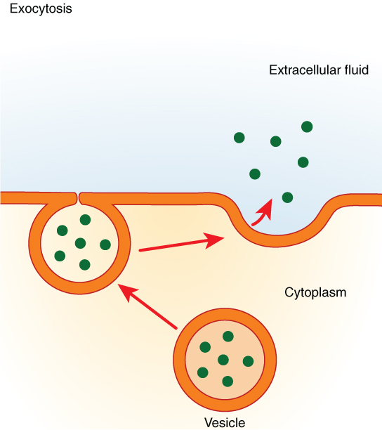
b) Exocytosis: Releasing Material Out
- How: A vesicle containing material fuses with the plasma membrane, releasing its contents outside the cell.
- Uses: Secreting hormones, neurotransmitters, digestive enzymes; removing cellular waste.

Summary
Membrane transport is vital for cellular life. Cells utilize:
- Passive Transport (Simple Diffusion, Facilitated Diffusion, Osmosis) to move substances down concentration gradients without energy expenditure.
- Active Transport (Primary and Secondary) to move substances against concentration gradients, requiring energy (ATP).
- Bulk Transport (Endocytosis, Exocytosis) to move large materials via vesicles, also requiring energy.
Ensure you understand the distinctions between these mechanisms, the role of membrane components (lipids, proteins), and the importance of concentration gradients and energy.
- Resources
- API
- Sponsorships
- Open Source
- Company
- xOperon.com
- Our team
- Careers
- 2025 xOperon.com
- Privacy Policy
- Terms of Use
- Report Issues
